Temporomandibular Joint Disease Treatment - North Shore Oral and Maxillofacial Surgery
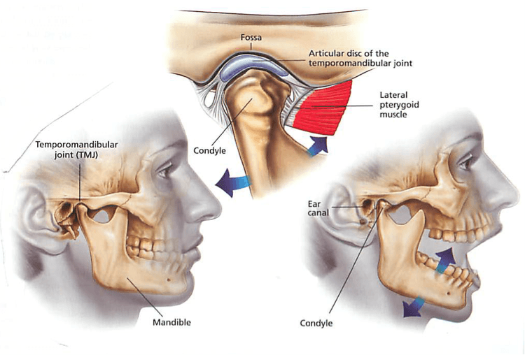
Temporo mandibular joint (TMJ) symptoms are frequent.
The most common is some clicking of the joint with mandibular movements, that in most cases is not painful.
Things change when pain with joint movement or biting and chewing occurs with or without clicking. This certainly indicates some sort of joint derangement, that needs attention.
Symptomatic treatment with anti-inflammatories can reduce the inflammation and related pain. This will help, but not solve the problem.
The underlying condition in many patients is a increased load of the joint through clenching or grinding of teeth. This often shows as soreness of the chewing muscles and stiffness in the morning. If those habits stay long enough, finally painful symptoms of the joints occur frequently.
If clenching or grinding of teeth is a contributing factor, it would be recommended to check, if any early teeth contacts may initiate the grinding. If that is not the case, usually an occlusal splint would be recommended. This is made from a model of the jaws, which is created from an impression of the jaw. This splint is placed on the upper or lower teeth as clear acrylic or soft appliance, that should be worn overnight in order to reduce any increased load on the joints. In case the patient grinds or clenches the teeth, this will no longer lead to increased load of the joints, as the splint maintains some distance between the teeth.
That way no pressure is created on the joint, even if the patient clenches or grinds the teeth. This splint as well protects the teeth from damage of grinding. This can otherwise lead to nerve irritation or cracked teeth and roots. This may lead to the need or root canal treatment or even the need for tooth removal, if there is a vertical tooth fracture occurring.
In addition to a splint the patient should have physiotherapy, reducing muscle hypertension by massage and giving exercise to strengthen the joint stabilising muscles. In many cases that will be sufficient to get the symptoms under control.
In case those measures would not lead to sufficient relief, further invasive treatment would have to be considered. The next step would be an arthrocentesis. This carried out under anaesthesia – usually iv sedation. With this procedure the joint is accessed with two needles, one for rinsing, the second one for drainage of the rinsing fluids. Additionally the joint is mobilised to get any debris detached and washed out. Finally, some Cortison is usually applied topically to reduce any inflammation.
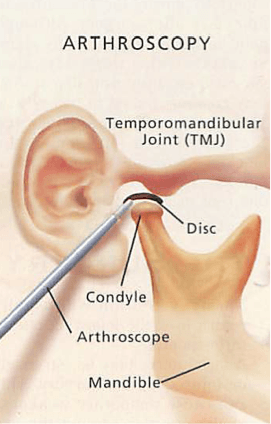
If this still would not fix the problem, the next step would be an arthroscopy. In addition to the arthrocentesis there would be a scope placed into the joint space to visualise the joint anatomy. That way further mobilisation and washout of debris could be achieved, which will usually improve the symptoms.